NON CARIOUS CERVICAL LESIONS
Tooth structure loss cannot be occur only by caries/cavities, Non carious cervical lesions also cause destruction of tooth substance at the cervical region [where the tooth and gum together-also called cement-enamel-junction].
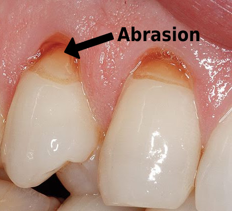
Abrasion:-
It is a non-carious cervical lesion,caused by mechanical wears of tooth from interaction with foreign [outside] objects other than tooth to tooth contact.

Etiology:-
- Most common cause is improper brushing method.
- Excessive frequency of brushing.
- More time and force applied during brushing.
- Using hard toothbrush and abrasive toothpaste .
- Using tooth powder - Tooth powder is generally more abrasive than tooth paste.
- Biting or Chewing on fingernails, pen tips or any other hard subjects.
- Ill fitting clasps of partial dentures.[removable dentures]
Abfraction:- It is pathological loss of tooth substance caused by bio mechanical loading forces that result in flexure and failure of enamel and dentin at a location away from loading.

Etiology-
- Using of excessive force on teeth while chewing or grinding.
- Habit of Bruxisum. [Involuntary grinding of teeth].
EROSION:-
Loss of tooth structure due to acids originated from internal [inside body] or from external source.

Etiology:-
A] Extrinsic/External Factors:
- Environmental/Occupational
:- Wine tasters, Swimmers
- Dietary
:- Soft drinks, Acidic drinks, Citrus fruits, Carbonated drinks
B] Intrinsic/Internal Factors:
- Gastric
disorders:
- Gastric
ulcers
- Hiatus
hernia
- Chronic
alcoholism
- Chronic vomiting
- Pregnancy
morning sickness
- Anorexia
nervosa
- Bulimia
nervosa
|
CLINICAL FEATURES
OF NON CARIOUS CERVICAL LESIONS
|
|
|
ABRASION
|
ABFRACTION
|
EROSION
|
|
LOCATION
|
Facial[outer surface]
|
Facial
|
Facial or Lingual[inner surface]
|
|
SHAPE
|
Wedged, Notched or Grooved
|
Wedge shape
|
Shallow “U” or disc shape
|
|
MARGINS
|
Sharp
|
Sharp
|
Smooth
|
|
ENAMEL SURFACE
|
Smooth, may show scratches
|
Initials stages: Rough, later may develop grooves
|
Smooth, may be polished
|
|
TEETH AFFECTED
|
Many teeth involve, usually
facial [outer side of tooth] surfaces of maxillary [upper jaw] left canine to
molar teeth in right hand person and vice versa.
|
Single toot involve, sub
gingival location
|
Many teeth involve, usually
lingual [inner side of tooth] surface
of maxillary anterior
|
When to check with Dentist:
- Teeth sensitivity
- Teeth pain
- Aesthetic problem
- Deep cervical lesions which has more prone to tooth fracture
- Cervical lesions which has more prone to cavities formation
- Infected gingiva[gums] due to caries or accumulation of plaque in the affected cervical lesion
Treatment Options: This treatment is recommended for those people who has very less exposure of dentin and experiences hypersensitivity. This is managed by following methods:
- Using desensitising toothpaste
- Fluoride varnish
- Dentin-bonding agents
If the affected area is deep/large, the lesion have prone to pulp expose, the person has more sensitivity, aesthetic problem, if caries started at the the cervical lesion, if the lesion seems to cause gingival infection
These lesions are managed by restoration. And most commonly using restorative materials are
- Composite resins
- Tooth coloured resins
- Glass inomer cement [GIC]
- Resin modified GIC and
- A combination of composite and GIC in a sandwich technique.
- Endodontic therapy:-
- When cervical lesion involves pulp exposing and leads to severe pain endodontic treatment [RCT-Root canal treatment] required.
- The tooth fractured at cervical region is indicated for endodontic treatment followed by post placement and crown preparation.
- Periodontal Therapy:-
- When Cervical lesions cause severe gingival infection due to more plaque accumulation at that region, sometime it leads to periodontal disease/Infection.
- Based on severity and affected area of gingiva it requires root coverage procedures using grafting and non grafting methods.


Comments
Post a Comment